
Caenorhabditis Elegans: The Tiny Worm Transforming Science
How a transparent, one-millimeter nematode became the only organism with a completely mapped nervous system, and what its 302 neurons are teaching scientists about memory, aging, and consciousness itself.
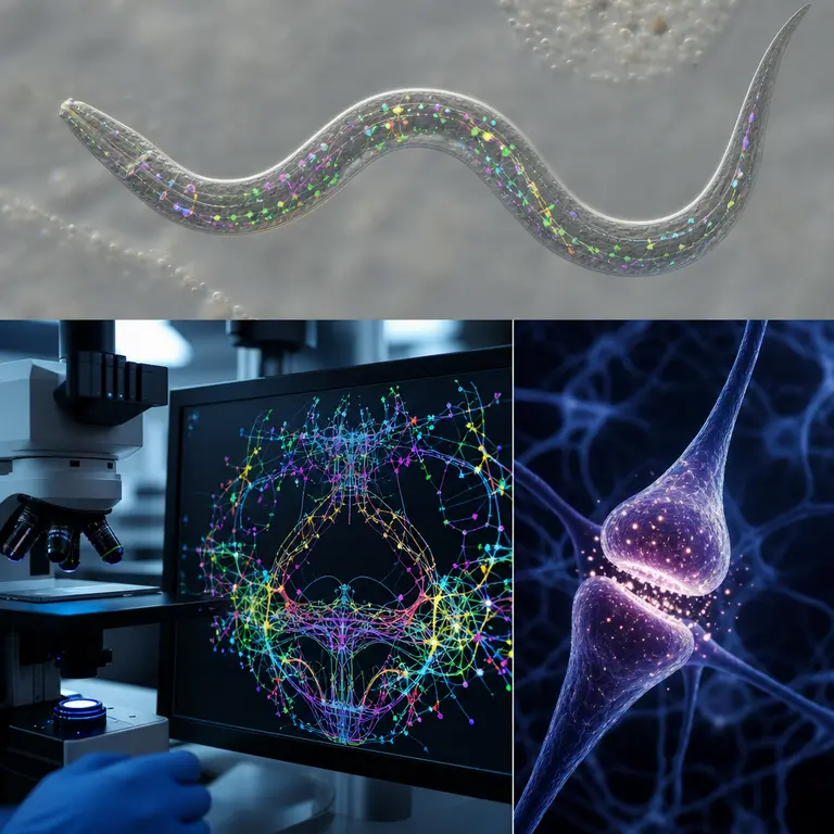
The Most Studied Animal You’ve Never Heard Of
In 1963, South African biologist Sydney Brenner made a decision that would seem absurd to most of his colleagues: he chose to dedicate his career to studying a nearly invisible, soil-dwelling roundworm with no eyes, no ears, and a brain containing exactly 302 neurons. The worm was Caenorhabditis elegans, and Brenner’s gamble eventually earned him a Nobel Prize in 2002. Today, C. elegans holds a distinction no other animal on Earth can claim — it is the only multicellular organism whose complete nervous system has been mapped, neuron by neuron, synapse by synapse, into a full wiring diagram called a connectome.
That connectome contains 302 neurons and exactly 7,000 synaptic connections. For comparison, the human brain contains approximately 86 billion neurons and an estimated 100 trillion synaptic connections. Yet the gap in complexity has not diminished C. elegans’s scientific value. If anything, its radical simplicity is precisely why it has become the most powerful model organism in modern neuroscience. The story of how a microscopic worm became the cornerstone of some of the most consequential biological discoveries of the twentieth and twenty-first centuries is one of the stranger and more instructive narratives in the history of science.
A Body Plan of Stunning Precision
C. elegans is transparent throughout its entire life cycle, which means researchers can watch every cell division, every nerve signal, and every developmental event in a living animal under a standard light microscope without dissection or staining. This single physical property — total optical transparency — transformed it from an obscure soil organism into a window onto the fundamental machinery of animal life. The adult hermaphrodite, the most commonly studied form of the species, contains exactly 959 somatic cells every time, in every individual, regardless of environmental conditions or geographic origin. This near-perfect cellular determinism is called eutely, and it is extraordinarily rare in the animal kingdom.
John Sulston, working alongside Brenner in Cambridge during the 1970s and 1980s, painstakingly traced the lineage of every one of those 959 cells from the fertilized egg forward, producing the first complete cell lineage map of any animal. The work required years of continuous observation, with Sulston sitting at a microscope for hours each day, manually recording each cell division as it occurred in real time. He discovered something startling in the process: 131 cells are programmed to die during normal development through a process called apoptosis, or programmed cell death. This was not damage or disease — it was deliberate biological demolition, built into the organism’s developmental plan from the moment of conception.
Sulston’s work revealed apoptosis as a fundamental mechanism of life, one that operates in virtually every multicellular organism on the planet, including humans. During human embryonic development, apoptosis sculpts fingers by eliminating the webbing between them, prunes excess neurons in the developing brain, and eliminates potentially cancerous cells before they can proliferate. Dysregulation of this process underlies many cancers, in which cells that should die instead survive and multiply uncontrollably. The discovery that programmed cell death was not an accident but a conserved and essential biological program was one of the most significant conceptual shifts in twentieth-century biology. Sulston shared the 2002 Nobel Prize with Brenner and fellow researcher Robert Horvitz, who had mapped the specific genes governing which cells lived and which died.
The Connectome and the Dream of Whole-Brain Simulation
The complete neural wiring diagram of C. elegans, published in its first form by White, Southgate, Thomson, and Brenner in 1986 in the journal Philosophical Transactions of the Royal Society B, was assembled from thousands of electron microscope images of cross-sectioned worm tissue. The project took over a decade of manual labor. Each image had to be examined by hand, each synapse identified and recorded, each connection traced across sequential tissue slices like a three-dimensional puzzle assembled one paper-thin layer at a time. The resulting map, known as the C. elegans connectome, remained the only complete connectome of any nervous system for nearly three decades, a testament both to the pioneering nature of the achievement and to the immense difficulty of replicating it in any more complex organism.
In 2012, a project called OpenWorm launched with the goal of simulating the entire organism in software — not just its neurons, but its muscles, body physics, and environmental interactions. The project brought together computational biologists, neuroscientists, and software engineers from multiple countries in an open-source collaboration that framed the worm as a biological engineering problem to be solved collectively. By 2014, researchers had uploaded the C. elegans connectome into a simple Lego robot body. Without any explicit programming for locomotion, the robot began to exhibit worm-like movement, responding to touch stimuli in ways that mirrored those of a living animal. The experiment was controversial and its interpretation vigorously debated, but it raised a question that philosophers and neuroscientists are still wrestling with: if you faithfully replicate a connectome in hardware, does something meaningful about the original organism transfer with it?
The Human Connectome Project, launched by the National Institutes of Health in 2009 with a $40 million budget, draws a direct intellectual lineage from the C. elegans work. The ambition to map the human brain’s wiring at full resolution remains computationally intractable for now — a single cubic millimeter of human cortex contains roughly 57,000 neurons and 150 million synapses, and the brain contains approximately 1.3 million cubic millimeters of tissue. The data storage requirements alone for a complete human connectome at synaptic resolution have been estimated at around one zettabyte, roughly equivalent to the entire current digital output of humanity. Nevertheless, C. elegans established a proof of concept that such mapping is possible and scientifically transformative, and advances in electron microscopy and machine-learning image analysis are steadily closing the gap between ambition and capability.
Aging, Longevity, and the Genes We Share
Perhaps the most medically significant contribution of C. elegans research has come from the field of aging biology. In 1993, Cynthia Kenyon at the University of California, San Francisco, discovered that a single mutation in a gene called daf-2 doubled the lifespan of C. elegans. The worm normally lives about three weeks; mutants with the daf-2 alteration lived six weeks or more, remained vigorous and active well into old age, and showed remarkable resistance to heat and oxidative stress. Kenyon reportedly described the mutant worms as moving through their extended lives like teenagers rather than the elderly, maintaining youthful activity levels far beyond the point at which normal worms had already died.
Daf-2 encodes a receptor for insulin-like growth factor signaling — a pathway that is conserved across virtually all animals, including humans. This conversation is not superficial. The molecular architecture of the pathway, the downstream signaling cascades it activates, and the cellular processes it regulates are recognizably similar across worms, flies, mice, and people. Kenyon’s discovery ignited the modern biology of aging and directly inspired pharmaceutical research into compounds that modulate the insulin and IGF-1 pathway in humans. Subsequent work in C. elegans identified dozens of longevity genes, many of which have human orthologs implicated in age-related diseases, including Alzheimer’s disease, Parkinson’s disease, and type 2 diabetes.
A 2003 study extended the record further: worms carrying mutations in both daf-2 and another gene called clk-1 lived up to five times their normal lifespan — the equivalent, scaled to human terms, of a person living to approximately 400 years. These worms did not simply survive longer in a diminished state; they remained functional and reproductively active for most of their extended lives, suggesting that the mutations compressed the period of decline rather than merely prolonged it. While direct translation to human longevity remains distant, and the biology of aging in mammals involves layers of complexity not present in a simple roundworm, these findings have fundamentally altered how scientists think about aging itself — as a genetically regulated, and therefore potentially modifiable, process rather than an inevitable entropic collapse that medicine can only slow but never redirect.
A Worm in Space and the Future of Neuroscience
C. elegans has even traveled to space, and done so under circumstances that no experimenter planned. In 2003, a culture of worms survived the catastrophic disintegration of the Space Shuttle Columbia during reentry and recovered alive from the debris field scattered across eastern Texas. The worms had been housed in a sealed container as part of a legitimate microgravity experiment, and the container’s insulating properties apparently protected them from the heat and impact of the breakup. NASA has since deliberately sent C. elegans to the International Space Station multiple times to study how microgravity affects muscle atrophy and gene expression — research with direct implications for human astronaut health on long-duration missions to the Moon, Mars, and beyond.
More recently, C. elegans has become a test bed for optogenetics, a technique in which individual neurons are genetically engineered to express light-sensitive proteins that allow them to be activated or silenced by pulses of light at specific wavelengths. Because the worm is transparent, researchers can target any specific neuron in a living, behaving animal with millisecond precision using a laser, then watch in real time how behavior changes as individual circuit elements are switched on or off. This has allowed scientists to decode the neural circuits underlying hunger, avoidance responses, mate-seeking, and even rudimentary forms of learning and memory in an intact, freely moving animal. The precision available in C. elegans optogenetics experiments is simply not achievable in any vertebrate system, where tissue opacity and cellular density make single-neuron targeting in a behaving animal extraordinarily difficult.
The worm that Brenner chose in 1963 has now been the subject of over 25,000 published scientific papers. It has contributed to six Nobel Prizes either directly or through discoveries it enabled. It has reshaped our understanding of development, cell death, aging, neural circuit function, and the very nature of mapping a mind. It remains, by nearly every measure, the most completely understood animal on the planet — and yet researchers continue to find surprises in its 302-neuron brain, new behaviors, new mechanisms, new questions that its apparent simplicity had concealed. The lesson may be that simplicity and depth are not opposites, and that the most productive scientific choices are sometimes the ones that look, to everyone else in the room, like the strangest ones imaginable.
Sources & Further Reading
- Brenner, Sydney. The Genetics of Caenorhabditis elegans. Genetics, 1974. https://www.genetics.org/content/77/1/71
- White, J.G., Southgate, E., Thomson, J.N., and Brenner, S. The Structure of the Nervous System of the Nematode Caenorhabditis elegans. Philosophical Transactions of the Royal Society B, 1986. https://doi.org/10.1098/rstb.1986.0056
- Kenyon, C., Chang, J., Gensch, E., Rudner, A., and Tabtiang, R. A C. elegans mutant that lives twice as long as wild type. Nature, 1993. https://doi.org/10.1038/366461a0
- OpenWorm Foundation. OpenWorm Project Documentation and Publications. openworm.org, 2014. https://www.openworm.org