
Cuttlefish: Colorblind Camouflage Masters with Skin Vision
Cuttlefish and octopuses don't just change color — they manipulate polarized light invisible to humans, communicating in a spectrum science is only beginning to decode.
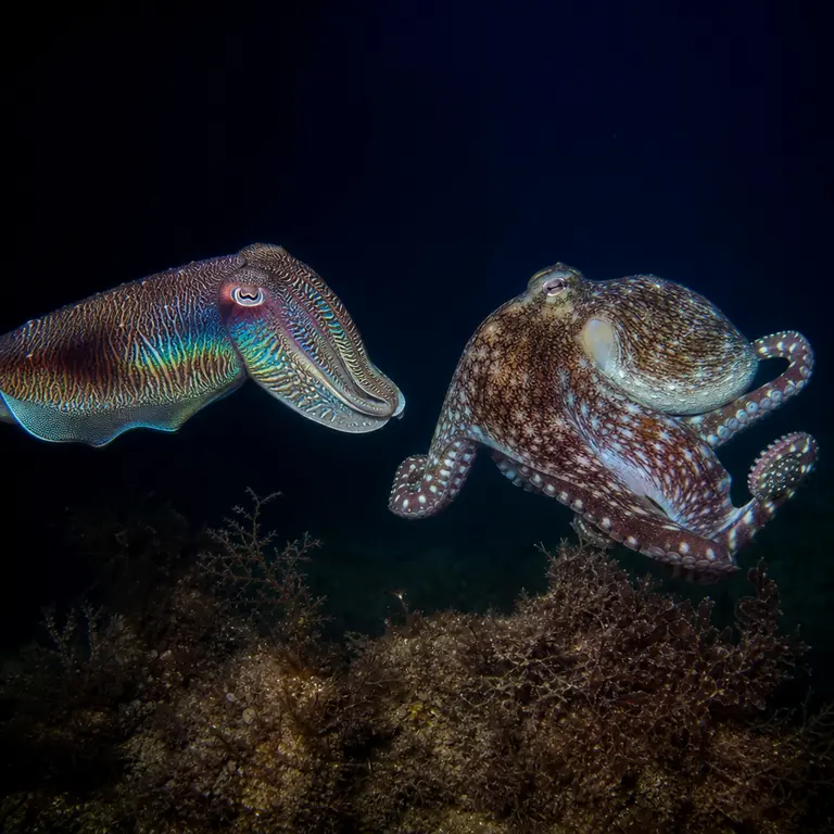
The Skin That Sees Without Eyes
Cuttlefish have no color-sensitive cone cells in their eyes. They are, by every conventional measure, colorblind. Yet they produce some of the most sophisticated and precisely color-matched camouflage displays in the animal kingdom, matching the hue, texture, and brightness of their surroundings with millisecond precision. This paradox has puzzled biologists for decades and remains one of the stranger unsolved problems in sensory neuroscience. To understand how a colorblind animal produces color-matched disguises, it is necessary to look not just at the eye, but at the skin, the nervous system, and the strange evolutionary logic that connects them.
How a Colorblind Eye Might Still Perceive Color
The leading hypothesis, first seriously proposed by Christopher Stubbs and colleagues at Harvard in 2010, suggests that cuttlefish may exploit chromatic aberration — the optical phenomenon in which different wavelengths of light focus at slightly different depths within the eye. By rapidly adjusting the size of their pupils, which are W-shaped and uniquely structured among all known animals, cuttlefish may effectively sample different focal planes and infer color information from what is essentially a monochromatic image. It is a workaround so elegant that it borders on the implausible, yet the mathematics supports it. The eye, in this model, is not a passive receiver but an active scanner, continuously sweeping through focal depths to reconstruct spectral information that its photoreceptors cannot directly detect.
This hypothesis remains unconfirmed, and the debate around it illustrates how difficult it is to study perception in animals that cannot report their own experience. Behavioral experiments have shown that cuttlefish can distinguish between colored targets in ways that suggest some form of wavelength discrimination, but isolating the mechanism responsible has proven elusive. Some researchers have proposed alternative explanations involving the spatial distribution of photoreceptors across the retina, or the possibility that skin photoreceptors contribute independently to the color-matching process. The skin itself, in some cephalopod species, contains light-sensitive proteins called opsins — the same class of molecules that form the basis of visual pigments in eyes. Whether these dermal opsins contribute to camouflage matching through a distributed, non-visual sensing system remains an open and genuinely exciting question. The possibility that an animal might see, in some functional sense, through its skin rather than its eyes is not a metaphor but a hypothesis under active investigation.
Chromatophores, Iridophores, and a Third Layer Nobody Expected
The skin of a cephalopod is not a single display system but a layered stack of distinct optical technologies operating simultaneously. The outermost layer contains chromatophores — pigment-filled sacs controlled by radial muscles that expand or contract within 200 milliseconds under direct neural command. Below them sit iridophores, which contain stacked plates of crystalline protein that reflect light through structural interference rather than pigment, producing iridescent greens, blues, and silvers that shift with viewing angle. Deepest of all lie leucophores, which scatter light broadly and provide a diffuse white background that amplifies the layers above. Together, these three systems interact in ways that no single layer could achieve alone, producing a display capable of matching brightness, color, texture, and spatial pattern simultaneously.
What researchers at the Marine Biological Laboratory in Woods Hole discovered in 2012, and what has since been confirmed through spectropolarimetric imaging, is that iridophores in several cephalopod species can be actively tuned by acetylcholine — a neurotransmitter more commonly associated with muscle contraction and memory formation in vertebrates. This means the structural color layer is not a passive optical filter but a dynamically adjustable one, capable of shifting its reflectance peak across tens of nanometers in under a second. The skin, in other words, is a programmable photonic crystal controlled by the same chemistry that fires synapses. The discovery was significant not only for what it revealed about cephalopod biology but because it demonstrated that structural color — long assumed to be fixed by geometry — could be brought under active physiological control. This principle has since become a target for materials scientists working on tunable photonic devices, with the cephalopod iridophore serving as a proof of concept that nature solved this engineering problem hundreds of millions of years ago.
The Polarized Light Channel Humans Cannot See
Beyond color and texture, cephalopods communicate through a channel entirely invisible to human observers: polarized light. While humans perceive light intensity and wavelength, we are essentially blind to its polarization — the directional oscillation of the electromagnetic wave. Most vertebrate predators share this limitation. Mantis shrimp represent the extreme end of polarization sensitivity, with 16 types of photoreceptors and the ability to detect circularly polarized light. Cuttlefish, though less extreme, possess photoreceptors oriented at right angles, giving them sensitivity to the plane of polarization that opens an entirely separate information channel layered on top of ordinary vision.
Research published in Current Biology by Lydia Mäthger and Roger Hanlon in 2006 demonstrated that cuttlefish display high-contrast polarization patterns on their arms during social interactions — patterns that are completely invisible against the background to any observer lacking polarization sensitivity, including most predatory fish. These hidden signals appear to serve as private communication channels, allowing cuttlefish to signal to conspecifics while maintaining camouflage from predators. It is the biological equivalent of a steganographic message: information concealed within an image that appears blank to the uninformed viewer. The iridophore layer is primarily responsible for generating these polarization signals, meaning the same skin structures that produce structural color also encode hidden social information. A single patch of cuttlefish skin is, simultaneously, a camouflage element, a color display, and a covert communication device — three functions operating in parallel through the same physical substrate.
Neurological Control at Extraordinary Speed
The speed and precision of cephalopod skin displays demand an extraordinary degree of neural organization. A single cuttlefish skin contains millions of chromatophores, each individually innervated by neurons running directly from the brain’s optic lobes. There is no intermediate processing layer, no hormonal delay — the display is essentially a direct neural projection onto the body surface. Researchers have described it as analogous to having a television screen wired directly to the visual cortex, bypassing every intermediate step. The resolution this achieves is remarkable: individual chromatophores can be activated in precise spatial sequences, producing moving waves, expanding rings, and complex textures that ripple across the body in patterns that appear almost deliberate in their choreography.
Work by Gilles Laurent’s group at the Max Planck Institute for Brain Research, published in 2018, used high-speed imaging during REM-like sleep states in cuttlefish and recorded rapid, dynamic changes in skin patterns that appeared to replay waking camouflage sequences. The animals, while apparently sleeping, cycled through flickering displays that resembled compressed versions of the patterns they had produced while hunting or hiding hours earlier. The implication — still contested — is that cephalopods may consolidate visual memory during sleep through skin replay, an idea that would make their integument not just an output device but part of the memory consolidation architecture itself. If true, it would represent a form of embodied memory with no known parallel in vertebrate neuroscience. The skin would not merely express what the brain decides, but would participate in the process by which experience becomes memory — a concept that challenges the assumption that cognition is confined to the brain.
Current Research and the Biomimicry Race
The practical implications of cephalopod skin architecture have not been lost on materials scientists and defense researchers. DARPA funded early-stage research into flexible, electronically controlled photonic skins as far back as 2012. More recently, a team at the University of California, Irvine, published a 2017 Science paper describing a soft, stretchable electroluminescent device inspired directly by cephalopod iridophore geometry, capable of changing color and pattern in real time. The device used a layered architecture of ionic conductors and dielectric elastomers that closely mirrored the biological stack of chromatophore, iridophore, and leucophore layers. Subsequent research groups have explored applications ranging from adaptive military camouflage to medical devices that change their optical properties in response to physiological signals.
None of these engineered systems yet approaches the resolution, speed, or energy efficiency of the biological original. A cuttlefish operates its entire display system on the metabolic equivalent of a dim LED, integrating sensing, computation, and output into a single continuous process with no clear boundary between where perception ends and expression begins. The gap between biological and engineered performance remains enormous, itself a measure of how far cephalopod evolution has traveled along an optical path that human engineering is only beginning to map.
Conclusion
The cuttlefish presents a sustained challenge to assumptions about how sensory systems must work. It achieves color matching without color vision, communicates without sound or visible signal, and may consolidate memory through a process distributed across its own skin. Each of these features, taken individually, would be remarkable. Together, they describe an organism whose entire body functions as an integrated sensory and expressive system, with no clear separation between input and output, perception and display. The deeper that researchers look into cephalopod neuroscience and optics, the more the conventional frameworks of sensory biology begin to strain. Whatever the cuttlefish is doing, it is doing it with a logic that evolution arrived at independently, along a path that diverged from vertebrate neuroscience more than five hundred million years ago. That such a path could lead to this degree of sophistication is, on its own, one of the more quietly astonishing facts in all of biology.
Sources & Further Reading
- Mäthger, L.M. and Hanlon, R.T. Functional iridescence from crystalline platelets in cephalopod chromatophores. Proceedings of the Royal Society B, 2007. https://royalsocietypublishing.org/doi/10.1098/rspb.2006.0240
- Stubbs, A.L. and Stubbs, C.W. Spectral discrimination in color blind animals via chromatic aberration and pupil shape. Proceedings of the National Academy of Sciences, 2016. https://www.pnas.org/doi/10.1073/pnas.1524578113
- Nadler, J.H. et al. Cuttlefish use startle displays, but not against large predators. Animal Behaviour, 2016.
- Gire, D.A. et al. Cuttlefish show active sleep-like states with rapid eye movements and skin pattern changes. Journal of Experimental Biology, 2019. https://journals.biologists.com/jeb/article/222/22/jeb195263/224166