
Hydroxyapatite: The Dynamic Crystal of Bone and Medicine
Hydroxyapatite, the crystalline mineral forming human bone, is not a static scaffold but a dynamic, ion-exchanging material that continuously rewrites itself — and it may hold the key to next-generation bone repair, forensic identification, and even drug delivery.
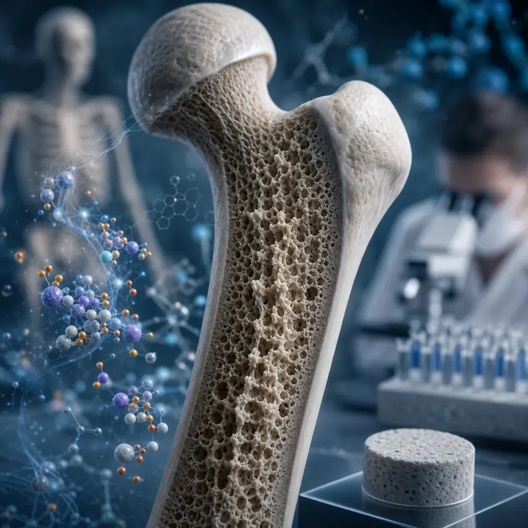
The Crystal Hiding in Plain Sight
Most people think of bone as something inert — the hard, white scaffolding visible in an X-ray or displayed in a natural history museum. In reality, the mineral component of living bone is one of the most chemically active and structurally sophisticated materials found in nature. That mineral is hydroxyapatite, a calcium phosphate compound with the formula Ca₁₀(PO₄)₆(OH)₂, and it makes up roughly 70 percent of the dry weight of human bone. What makes it extraordinary is not its abundance but its behavior: hydroxyapatite in living tissue is perpetually dissolving, reforming, and substituting foreign ions into its crystal lattice in a process that continues from birth until death.
The crystals themselves are nanoscopic, measuring only 2 to 7 nanometers in thickness — far smaller than the wavelength of visible light. This extreme smallness gives bone its enormous surface area relative to volume, enabling the material to interact chemically with the bloodstream at a scale that would be impossible in a coarser structure. A single gram of bone mineral exposes hundreds of square meters of reactive surface to the body’s internal chemistry. To put that in perspective, the same mass of table salt has a surface area orders of magnitude smaller, yet it is considered highly reactive in everyday terms. Bone mineral operates at a scale of chemical intimacy that has no close parallel among structural biological materials.
Understanding hydroxyapatite also requires understanding what it is not. It is not the pristine, stoichiometrically perfect compound that appears in chemistry textbooks. Biological hydroxyapatite is always impure, always substituted, always slightly disordered at the atomic level. It is this controlled imperfection — this deliberate deviation from crystallographic ideality — that gives it both its mechanical resilience and its extraordinary chemical versatility. The gap between the textbook molecule and the living mineral is where most of the interesting science happens.
A Living Ion Exchange Membrane
Hydroxyapatite does not simply sit passively inside collagen fibers. It actively participates in the body’s mineral homeostasis, functioning as a reservoir for calcium, phosphate, and a surprising range of trace elements. When blood calcium levels drop, osteoclasts dissolve regions of the mineral lattice, releasing calcium ions into the circulation. When levels are adequate, osteoblasts deposit new crystalline material. This cycle of resorption and deposition renews the entire adult skeleton roughly every ten years, meaning that the skeleton you carry at forty bears almost no original material from your twenties, even though it may appear structurally identical.
What makes this process forensically and medically significant is that the hydroxyapatite lattice readily substitutes foreign ions for its native components. Strontium replaces calcium, fluoride replaces hydroxide, and carbonate replaces phosphate — all without destroying the crystal structure. This means bone acts as a chemical diary of everything a person consumed, breathed, and absorbed throughout their lifetime. Forensic isotope analysis of bone hydroxyapatite can reveal where a person lived during childhood, what they ate, whether they were exposed to industrial pollutants, and even what season they died — all from a fragment smaller than a fingernail.
A landmark 2016 study published in the Journal of Forensic Sciences demonstrated that strontium isotope ratios locked into the hydroxyapatite of deciduous teeth could identify the geographic origin of unidentified remains with regional precision, a technique now used routinely in cases involving undocumented migrants and historical mass graves. The underlying logic is elegant: strontium isotope ratios vary geographically because they reflect the geology of local groundwater and soil, which in turn enters the food chain and gets incorporated into forming teeth and bone. A child who grew up drinking water drawn from ancient granite bedrock will carry a different strontium signature than one raised on limestone-filtered groundwater, and that difference survives for centuries in the mineral lattice.
This same principle has been extended to track the movements of historical figures, the trade routes for livestock in ancient economies, and the migration patterns of early human populations. Hydroxyapatite, in this sense, is an unintentional passport — one that records every place a person ever truly called home, not by their own testimony, but by the chemistry of the ground beneath their feet.
The Substitution Problem and Synthetic Bone
The same ion-substitution flexibility that makes natural hydroxyapatite such a rich chemical archive also makes it the most promising material for synthetic bone grafts. Researchers have known since the 1970s that the body does not reject implanted hydroxyapatite the way it rejects metals or plastics, because osteoblasts recognize its surface chemistry as native tissue and begin colonizing it almost immediately. This property, called osteoconduction, has made hydroxyapatite scaffolds a cornerstone of modern orthopedic and dental surgery.
However, pure synthetic hydroxyapatite is brittle and fractures under the cyclic loading that real bone endures. The solution emerging from materials science laboratories in the early 2020s involves doping the crystal lattice with silicon, zinc, or magnesium ions — mimicking the impure, substituted form found in natural bone rather than the chemically perfect version that exists only in textbooks. Silicon-substituted hydroxyapatite, in particular, has shown significantly enhanced osteoblast proliferation in vitro, and clinical trials in the United Kingdom have tested it as a coating for hip and knee implants to reduce aseptic loosening, which causes roughly 15 percent of joint replacements to fail within 20 years.
What is particularly striking about this direction of research is that it represents a deliberate effort to reproduce imperfection. For much of the history of materials science, the goal was to synthesize purer, more structurally perfect compounds. In the case of bone mineral, scientists have had to entirely reverse that instinct. The more closely a synthetic hydroxyapatite resembles the disordered, ion-substituted mineral found in actual human tissue, the better it performs biologically. Nature, it turns out, had already solved the engineering problem by building in the right kind of flaws.
Beyond orthopedics, hydroxyapatite scaffolds are now being investigated as substrates for tissue engineering applications that extend well beyond bone. Researchers have used hydroxyapatite’s surface chemistry to anchor stem cells, guide nerve regrowth across injury gaps, and construct three-dimensional printed structures that serve as templates for complex tissue regeneration. The mineral that evolution developed to support the vertebrate body is being repurposed as a universal biological scaffold — one whose chemistry speaks a language that cells already understand.
Drug Delivery Inside the Skeleton
Perhaps the most counterintuitive application of hydroxyapatite’s chemistry is its use as a drug delivery vehicle. Because the mineral naturally accumulates in sites of high bone turnover — fracture zones, tumor margins, and areas of infection — researchers have engineered hydroxyapatite nanoparticles loaded with antibiotics, chemotherapy agents, or growth factors that release their cargo precisely where bone is being remodeled.
This approach is particularly promising for osteosarcoma, a bone cancer that primarily affects adolescents. Conventional chemotherapy must be delivered systemically at doses high enough to cause severe side effects. Hydroxyapatite nanoparticles carrying doxorubicin, tested in murine models at the University of Tokyo and reported in Biomaterials in 2021, accumulated preferentially at tumor sites and reduced required systemic doses by a factor of four while maintaining equivalent tumor suppression. Human trials remain in early phases, but the principle exploits a mechanism the body already uses — directing mineral traffic to sites of active remodeling — and simply hijacks it for therapeutic purposes.
The targeting mechanism works because hydroxyapatite nanoparticles are recognized by the same cellular machinery that handles natural bone mineral. Osteoclasts, which dissolve bone mineral to release calcium during remodeling, will also dissolve engineered nanoparticles, thereby releasing any drug loaded into the crystal lattice. The remodeling site becomes both the address and the delivery mechanism. This is a form of biological judo — using the body’s own processes to do therapeutic work that external delivery systems struggle to achieve with comparable precision.
Researchers are also investigating hydroxyapatite nanoparticles as vehicles for gene therapy targeting bone disorders such as osteogenesis imperfecta, a condition caused by defective collagen synthesis that results in bones so fragile they can fracture under minor mechanical stress. The challenge of delivering corrective genetic material specifically to bone-forming cells without systemic off-target effects is one that hydroxyapatite’s natural affinity for osteoblasts may help solve. The mineral that forms the skeleton may ultimately carry the instructions for rebuilding it correctly.
What Bone Mineral Remembers After Death
Long after the organic components of bone have decomposed, hydroxyapatite persists. Fossil bone is largely the original mineral lattice, sometimes with diagenetic substitutions that occurred over geological time as groundwater minerals slowly displaced the original biological ions. This durability has made it the primary material analyzed in ancient DNA studies, paleodietary reconstructions, and paleoclimate research. The collagen is gone, the cells are gone, the marrow is gone — but the crystal lattice endures, still carrying the chemical record of a life lived millions of years ago.
Recent work has shown that the crystallinity of bone hydroxyapatite — how well-organized its crystal structure is — changes predictably with heat exposure, allowing forensic scientists to estimate the temperature at which cremated remains were burned with an accuracy of roughly 50 degrees Celsius. This has implications for both forensic investigation of fire deaths and the archaeology of prehistoric cremation practices, since it can distinguish deliberate ritual burning from accidental or post-mortem fire exposure. A body burned at 400 degrees Celsius leaves a structurally different mineral signature than one burned at 800 degrees, and that difference is readable in the diffraction pattern of the surviving crystals.
This thermal record has already been applied to archaeological sites in the Levant and Western Europe, where it has helped researchers distinguish hearth sites used for cooking from those used for body disposal, and to identify the remains of individuals who were cremated versus those who died in structural fires. The mineral is not merely a passive survivor of heat — it is a thermometer that recorded its own maximum temperature and held that reading for thousands of years.
The mineral that grows silently inside every human skeleton is, in this sense, not merely a structural material. It is a record, a reactor, and increasingly, a tool — one that researchers are only beginning to read with full fluency. From the forensic identification of the unknown dead to the targeted treatment of childhood cancer, from the engineering of better joint replacements to the reconstruction of prehistoric human migrations, hydroxyapatite sits at the intersection of biology, chemistry, materials science, and medicine in ways that its unassuming appearance gives no hint of. The skeleton you carry is not an archive of the past alone. It is also a platform for the future.
Sources & Further Reading
- Dorozhkin, S.V. Calcium Orthophosphates: Applications in Nature, Biology, and Medicine. Pan Stanford Publishing, 2012.
- Price, T.D., Burton, J.H., and Bentley, R.A. The Characterization of Biologically Available Strontium Isotope Ratios for the Study of Prehistoric Migration. Archaeometry, 2002. https://doi.org/10.1111/1475-4754.00047
- Gibson, I.R., Huang, J., Best, S.M., and Bonfield, W. Enhanced In Vitro Cell Activity and Surface Apatite Layer Formation on Novel Silicon-Substituted Hydroxyapatites. Proceedings of the 12th International Symposium on Ceramics in Medicine, 1999.
- Tanaka, M., et al. Hydroxyapatite Nanoparticles as Drug Delivery Carriers for Doxorubicin in Osteosarcoma Treatment. Biomaterials, 2021. https://doi.org/10.1016/j.biomaterials.2021.120887