
Krypton-79: a Forgotten Beacon in Medical Imaging History
Before MRI, a radioactive gas called krypton-79 was briefly used in the 1950s and 60s to visualize blood flow and lung function in living patients — a largely forgotten chapter in medical imaging history.
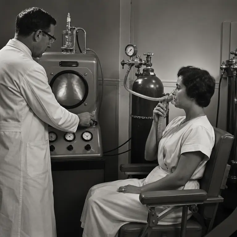
A Problem Without a Picture
For most of medical history, the interior of a living human body was inaccessible without a scalpel. X-rays, developed in 1895, could reveal bones and dense masses, but soft tissue — the lungs filling with air, blood coursing through vessels, the subtle collapse of a bronchial passage — remained essentially invisible. Physicians could observe a patient struggling to breathe, measure the crude volumes of air moved in and out of the chest, and listen through a stethoscope for sounds that hinted at obstruction or fluid. What they could not do was see where, within the lung's architecture, the problem actually lived.
By the 1940s and 1950s, physicians working in cardiology and pulmonology were acutely aware of this gap. Diagnosing conditions like pulmonary embolism, congenital heart defects, or regional lung disease required inference from symptoms, crude measurements, and the occasional exploratory procedure that carried real risk. Pulmonary angiography, for instance, involved threading a catheter into the heart and injecting contrast dye into the pulmonary arteries — a procedure that occasionally caused the very catastrophe it was meant to diagnose. The body’s functional geography was largely a matter of educated guesswork, and the guesses sometimes killed people.
The solution that briefly emerged from this frustration was not the one history ultimately remembered. Before the now-ubiquitous technetium-99m scan, before positron emission tomography, and long before magnetic resonance imaging, a small cohort of researchers in Britain and the United States turned to an unusual candidate: krypton-79, a radioactive isotope of the noble gas krypton, with a half-life of approximately 34.9 hours. The story of how they used it, why it worked, and why it disappeared is a window into a period of medical experimentation that has been almost entirely absorbed into the footnotes of radiology history.
Krypton-79 and the Logic of Inert Gases
The appeal of krypton-79 was rooted in chemistry — or more precisely, in the near-total absence of it. Noble gases do not react with biological tissue. They do not bind to proteins, alter cell membranes, or trigger immune responses. Inhaled or injected into the bloodstream as a dissolved gas, krypton-79 would travel passively wherever blood or air carried it, emitting detectable gamma radiation as it decayed, and then disappear from the body without leaving a chemical trace. For a medical community that had watched patients suffer allergic reactions to iodine-based contrast agents and endure the cumulative effects of repeated radiation exposures, a tracer that was both inert and self-eliminating was a genuinely attractive proposition.
Researchers at Hammersmith Hospital in London, particularly the team associated with the Medical Research Council, began exploring radioactive noble gases for physiological measurement in the late 1950s. The physicist-physiologist collaboration that drove this work was characteristic of the era: physicists who had cut their teeth on wartime isotope production were looking for peacetime applications, while clinicians were desperate for non-destructive ways to measure function rather than just structure. The two communities found each other in the corridors of institutions that had been reorganized by wartime science and were now flush with government funding directed toward civilian benefit.
Krypton-79 was produced in cyclotrons by bombarding stable bromine-79 with protons. The resulting isotope emitted positrons and gamma rays at energies that early scintillation detectors could capture. When a patient inhaled krypton-79 mixed with oxygen, the gas distributed through the lungs in proportion to regional ventilation — areas receiving more airflow accumulated more tracer. When injected intravenously as a saline solution equilibrated with the gas, it followed perfusion patterns, accumulating in well-supplied tissues and bypassing blocked or poorly vascularized regions. The principle was elegant in its simplicity: the gas went where the body sent it, and the radiation told you where that was.
The resulting scans, printed as crude maps of radiation counts across the chest or abdomen, could distinguish a functioning lung lobe from a collapsed one, or reveal the distribution asymmetry caused by a pulmonary embolism. Researchers could observe that one region of a patient’s lung was receiving adequate airflow but inadequate blood supply — the hallmark pattern of an embolism — or that a child’s heart was shunting blood in the wrong direction through a septal defect. For the mid-1950s, this was remarkable. For the first time, a physician could look at a map of function rather than a shadow of structure.
Clinical Use and Its Limits
Between roughly 1956 and 1968, krypton-79 scanning was used in a modest number of clinical research settings, primarily in the United Kingdom, Sweden, and at a handful of American academic medical centers. Published studies appeared in journals including Clinical Science and the British Journal of Radiology. Researchers used the technique to study regional lung ventilation in patients with emphysema, to assess cardiac shunts in children with congenital defects, and to map cerebral blood flow in experimental settings. The volume of published work was never large — perhaps several dozen substantive papers over the course of a decade — but the quality of the physiological reasoning in those papers was often sophisticated, anticipating questions that would not be answered better for another twenty years.
The technique had genuine advantages. The short biological retention time — noble gases are rapidly exhaled or diffuse out of tissue — meant that radiation doses were relatively low compared with some contemporary contrast agents. The chemical inertness eliminated concerns about toxicity or allergic reaction. And the ability to repeat measurements within hours, owing to rapid clearance, allowed researchers to observe dynamic physiological changes rather than a single frozen moment. A patient could be scanned before and after a bronchodilator, or before and after a change in body position, and the results could be compared with some confidence that the tracer itself had not altered the measurement.
But krypton-79 also had serious practical limitations that accumulated into an insurmountable disadvantage. Cyclotron production was expensive and geographically constrained — only institutions near a particle accelerator had reliable access, and in the late 1950s, those institutions were few. The 34.9-hour half-life was long enough to complicate waste handling but short enough to create logistical headaches for shipping; a batch produced on Monday might be barely usable by Wednesday and entirely expired by the following week. The scintillation cameras of the era produced low-resolution images by any modern standard, and interpreting the count maps required considerable expertise in both physics and anatomy. A bright spot on the scan could represent genuine tracer accumulation, a detector artifact, or scattered radiation from an adjacent structure, and distinguishing between these possibilities demanded a level of technical fluency that most clinical departments could not sustain.
When technetium-99m became widely available through the molybdenum-99 generator system in the mid-1960s — a technology that allowed hospitals to produce their own fresh isotope on-site without a cyclotron — the economics changed decisively. Technetium-99m offered superior gamma-ray energy for imaging, a convenient six-hour half-life, and the ability to be chemically attached to a vast range of targeting molecules, making it adaptable to virtually every organ system. A hospital could install a generator, receive weekly shipments of the parent isotope molybdenum-99, and produce fresh technetium doses on demand. No cyclotron, no physicist, no specialized infrastructure beyond a basic nuclear medicine suite. Krypton-79 scanning quietly faded from clinical practice without ever having been formally abandoned. It was not proven wrong so much as rendered obsolete by a more versatile and more accessible competitor.
The Broader Noble Gas Moment
Krypton-79 was not the only radioactive noble gas explored during this period, and its story makes more sense when placed within the wider landscape of mid-century isotope medicine. Xenon-133, with a half-life of 5.2 days, proved more durable in clinical practice and remained in use for lung ventilation studies well into the 1990s, particularly in nuclear medicine departments that had already invested in the infrastructure to handle gaseous tracers. Krypton-81m, with a half-life of just 13 seconds, was later developed as an ultra-short-lived ventilation agent that could be continuously inhaled during a scan, allowing real-time imaging of airflow distribution without significant radiation accumulation. Radon-222 was briefly investigated but quickly abandoned due to its toxicity as a decay product of radium and its well-documented association with lung cancer risk — an irony that would have been grimly apparent to any pulmonologist.
The krypton-79 episode sits within a broader pattern of mid-twentieth-century radioisotope medicine that is now largely invisible to practitioners trained after the MRI era. The 1950s and 1960s were a period of extraordinary isotope experimentation, driven partly by the political imperative to demonstrate peaceful uses for nuclear technology in the aftermath of Hiroshima and Nagasaki. Government funding in the United States, channeled through the Atomic Energy Commission, and in Britain through the Medical Research Council, actively encouraged clinicians to find medical applications for cyclotron and reactor byproducts. Isotopes were available, the infrastructure to produce them existed, and there was genuine institutional pressure to show that the same science that had built atomic weapons could also heal. Some of these experiments were conducted with insufficient attention to patient consent — a shadow that hangs over the entire era of radioisotope clinical research, and one that subsequent historians have documented with uncomfortable specificity.
A Present-Day Echo in Hyperpolarized Gas Imaging
The logic that made krypton-79 attractive in 1958 has not disappeared — it has been refined into one of the most promising frontiers in pulmonary imaging today. Hyperpolarized noble gas MRI, which uses krypton-83 or helium-3 that has been magnetically polarized to enhance MRI signal by a factor of up to 100,000, allows clinicians to visualize airflow distribution in the lungs with extraordinary spatial resolution and no ionizing radiation whatsoever. The technique exploits the same fundamental property that drew researchers to krypton-79 seven decades ago: an inert gas inhaled into the lungs will distribute according to the mechanics of ventilation, and if you can detect where it goes, you can map how the lung is actually functioning rather than merely what it looks like on a structural image.
In 2023 and 2024, research groups at the University of Sheffield and at institutions in Canada and the United States published results demonstrating that hyperpolarized krypton-83 MRI can detect early structural and functional changes in the lungs of patients with long COVID, asthma, and chronic obstructive pulmonary disease at a stage when conventional CT scans appear entirely normal. The technique is moving toward clinical trials for regulatory approval in several countries. A 2024 review in the journal Radiology highlighted hyperpolarized gas MRI as one of the most significant emerging tools in functional lung imaging, with particular promise for pediatric patients who are most vulnerable to cumulative radiation exposure from repeated conventional scans. For children with cystic fibrosis who require monitoring throughout their lives, a technique that provides superior functional information without any radiation dose is not merely a technical improvement — it is a meaningful reduction in lifetime risk.
The continuity is striking and, on reflection, not entirely coincidental. The same element, the same physiological logic — inert gas follows airflow, distribution reveals function — and the same clinical question about regional ventilation that occupied researchers at Hammersmith in 1959 now occupies researchers at Sheffield and Toronto. The technology surrounding the gas has been transformed almost beyond recognition, from crude scintillation counts to three-dimensional MRI volumes acquired in a single breath-hold. But the underlying insight has persisted across seven decades, carried forward not by a continuous research tradition but by the independent rediscovery of a principle that was always sound.
What the Forgotten Technique Reveals
The history of krypton-79 scanning offers a useful corrective to the way medical progress is usually narrated. The story of imaging is typically told as a sequence of landmark inventions: X-ray, ultrasound, CT, MRI, PET. Each appears as a discrete breakthrough, attributed to a named inventor, celebrated with a Nobel Prize or a commemorative lecture. What this narrative obscures is the dense undergrowth of partial solutions, dead ends, and techniques that worked well enough to be used but not well enough to survive competition — and the researchers who built genuine knowledge within those constraints, often without recognition that extended beyond their immediate field.
The physiologists who worked with krypton-79 were not wrong about the principle. They were working at the edge of what their instrumentation and isotope supply chains could support, and they produced real clinical insight about lung disease and cardiac function that informed later work, even when that later work did not cite them directly. The fact that their specific tool was superseded does not diminish the quality of their reasoning. Science progresses not only through the accumulation of correct answers but through the refinement of good questions, and the question these researchers were asking — how do we see function rather than structure, how do we map the living physiology of a breathing, circulating body — turned out to be one of the most generative questions in twentieth-century medicine.
If anything, the reappearance of krypton in the hyperpolarized MRI literature suggests that the original intuition was more durable than the technology that first attempted to act on it. The researchers at Hammersmith did not fail to understand the lung. They succeeded in understanding it, using the only tools their era offered, and then watched those tools become obsolete. Medical history is full of such episodes: techniques that were genuinely useful, genuinely limited, and genuinely forgotten — waiting, in some cases, for a different era’s instruments to make the underlying idea viable again. Krypton-79 is one of the cleaner examples, precisely because the idea it embodied has now returned, wearing the considerably more sophisticated clothing of modern magnetic resonance physics, to answer the same questions its inventors first thought to ask.
Sources & Further Reading
- West, J.B. 'Historical aspects of the early Soviet/Russian manned space program.' Journal of Applied Physiology, 2001.
- Okpara, U.O. et al. 'Hyperpolarized noble gas MRI of the lung: current status and future directions.' Radiology, 2024. https://pubs.rsna.org/journal/radiology
- Dolphin, G.W. and Eve, I.S. 'The use of radioactive gases in assessing lung function.' Physics in Medicine and Biology, 1963.
- Chapman, S. et al. 'Hyperpolarized 129Xe and 83Kr MRI for pulmonary functional imaging.' NMR in Biomedicine, 2014.