
The Bone That Grows Back: Deer Antler Regeneration
Deer antlers are the fastest-growing tissue in the mammalian kingdom and the only known case of complete organ regeneration in adult mammals — a biological anomaly now driving serious cancer and regenerative medicine research.
 Watch on YouTube
 Watch on Rumble
The Only Organ a Mammal Can Regrow
Every spring, across the forests of North America, Europe, and Asia, male deer perform one of biology’s most astonishing feats: they grow an entirely new set of antlers from scratch. These structures, which can reach lengths of over 1.2 meters in elk and weigh up to 18 kilograms in moose, are not horns made of keratin, as in cattle or rhinoceroses. They are true bone — vascularized, mineralized, and neurologically innervated — and they regrow each year completely after being shed. No other adult mammal regrows an entire organ at this speed and with this regularity. The closest biological parallel in vertebrates is limb regeneration in axolotls, an amphibian that operates under entirely different developmental rules.
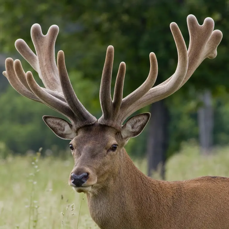
The growth rate alone is extraordinary. At peak season, antlers can grow as fast as 2.5 centimeters per day, making them the fastest-growing tissue ever recorded in any mammal. During this phase, the antlers are covered in velvet, a soft skin dense with blood vessels, nerves, and stem cells that supply the raw materials for mineralization. When antler growth is complete, the velvet dries and sheds, leaving the hardened bone used in combat and display. The entire cycle — from bare pedicle to full antler to shedding — takes roughly a year and is tightly governed by photoperiod, the seasonal change in day length, which is mediated by melatonin and testosterone signaling.
What makes this process even more remarkable is the sheer metabolic cost it imposes on the animal. During peak antler growth, a bull elk may redirect a significant portion of its daily caloric intake toward building bone tissue at a rate that has no equivalent anywhere in the mammalian kingdom. Calcium and phosphorus are mobilized from the skeleton itself during periods of nutritional stress, meaning the animal is essentially cannibalizing its own bones to build new ones. Despite this, the pedicles — the permanent bony platforms from which antlers emerge — remain structurally intact year after year, suggesting a level of tissue management and mineral regulation that physiologists are still working to fully understand. The deer is, in a very real sense, running a biological construction project that would exhaust the resources of most other large mammals within weeks.
The Stem Cell Secret Buried in the Skull
The biological engine behind this regeneration is a population of cells at the base of the antler, in a structure called the pedicle, a permanent bony protrusion from the skull. These cells, known as antler stem cells or antlerogenic periosteum cells, behave unlike virtually any other mammalian stem cell population. They retain an almost embryonic potency throughout the animal’s adult life, capable of initiating de novo bone formation season after season without exhausting their regenerative capacity or accumulating the genetic errors that typically accompany rapid cell division in other tissues.
Researchers at Shandong University in China published a landmark study in 2019 in the journal Science identifying a specific gene, TWIST2, as a key regulator of antler stem cell identity. When the team transplanted antlerogenic periosteum cells to other locations on a deer’s body, those cells initiated new antler growth at the transplant site — demonstrating that the regenerative signal is intrinsic to the cell population, not merely a response to local skull anatomy. This finding has profound implications for regenerative medicine because it suggests that a stable, self-renewing stem cell population capable of building complex mineralized tissue might be replicable or transferable in therapeutic contexts.
What distinguishes these cells from other stem cell populations studied in mammals is their apparent immunity to replicative senescence, the process by which cells progressively lose their ability to divide accurately as they age. Most rapidly dividing cells in adult mammals accumulate telomere shortening and DNA damage over time, eventually triggering programmed cell death or malignant transformation. Antlerogenic periosteum cells appear to circumvent this limitation through mechanisms that researchers have not yet fully characterized, but which likely involve unusually active DNA repair pathways and tightly regulated epigenetic controls. Understanding how these cells maintain their potency across decades of repeated activation could reframe how biologists think about the relationship between aging, stem cell exhaustion, and tissue repair capacity in mammals more broadly.
The pedicle itself is also a biological anomaly. Unlike most bone tissue, which forms during embryonic development and remains largely static in adulthood, the pedicle is a structure that first appears at puberty under the influence of androgens and then serves as a permanent launch platform for annual regeneration. It is, in effect, a specialized organ whose sole purpose is to house and protect the stem cell population that drives antler growth. No analogous structure has been identified in any other mammal, and its evolutionary origins remain a subject of active debate among paleontologists and developmental biologists.
Antlers, Cancer, and the Paradox of Controlled Growth
The rate at which antler tissue grows should, by ordinary biological logic, produce tumors. Rapid, sustained cell proliferation is precisely the hallmark of cancer, and the molecular pathways that drive antler growth — including IGF-1, Wnt signaling, and VEGF-mediated vascularization — are the same pathways implicated in some of the most aggressive human cancers. Yet antlers do not become malignant. The tissue grows, mineralizes, and stops with extraordinary precision, then is shed, and the cycle begins again.
This paradox has attracted the attention of oncologists and cell biologists who seek to understand the molecular brakes that prevent antler tissue from entering uncontrolled proliferation. One hypothesis involves the tight hormonal regulation of the cycle: testosterone surges in autumn trigger mineralization and velvet shedding, effectively halting growth by cutting off blood supply to the antler surface. Another hypothesis focuses on the velvet's microenvironment, which appears to produce a suite of anti-angiogenic and apoptotic signals that coordinate the shutdown of growth at precisely the right moment. A 2021 study published in Science identified that deer genomes contain expanded families of tumor suppressor genes compared to other mammals, suggesting that the capacity for rapid growth co-evolved with enhanced cancer resistance at the genomic level.
The implications of this finding extend well beyond deer biology. If the evolution of rapid tissue growth in cervids required a simultaneous expansion of cancer-suppression mechanisms, this suggests that growth and restraint are not opposites in the biological sense but are instead deeply coupled processes that must evolve together. This challenges a common assumption in oncology, which tends to treat cancer suppression as a passive or default state that breaks down under mutational pressure. In deer, suppression appears to be an active, elaborate, and dynamically regulated system that scales with the growth demands placed on the tissue. Identifying the specific tumor suppressor genes involved and understanding how they are activated in coordination with growth signals could offer a new conceptual framework for cancer prevention research in humans.
There is also an intriguing evolutionary dimension to this story. Antlers are a relatively recent innovation in mammalian evolution, appearing in the fossil record roughly 30 million years ago in early cervids. The co-evolution of rapid bone growth and expanded cancer resistance over that time period represents a compressed and observable example of how selection pressure can drive the simultaneous development of seemingly contradictory traits. Deer are, in this sense, a living experiment in evolutionary oncology — a field that examines how different species have solved the problem of preventing cancer under varying biological constraints.
From Forest to Laboratory: Therapeutic Frontiers
Interest in antler biology among medical professionals is not new. In traditional Chinese and Korean medicine, velvet antler has been used for over 2,000 years to treat joint pain, fatigue, and sexual dysfunction. Modern pharmacological analysis has confirmed that velvet antler contains insulin-like growth factor 1 (IGF-1), collagen precursors, chondroitin sulfate, and a range of bioactive peptides. While many traditional claims remain unsupported by rigorous clinical trials, the biological activity of velvet antler extracts in stimulating cartilage repair in animal models has been replicated across multiple peer-reviewed studies.
More compelling is the emerging research into antler-derived signals for bone repair in humans. Osteoporosis affects over 200 million people worldwide, and current treatments largely slow bone loss rather than stimulate meaningful new bone formation. The antlerogenic periosteal cells studied by the Shandong team represent a potential model for understanding how to instruct adult mammalian cells to rapidly and in large quantities build bone. Separately, researchers at the University of Georgia have investigated whether the unique extracellular matrix proteins expressed during velvet growth could be adapted into scaffolding materials for bone and cartilage tissue engineering. The field remains early-stage, but the deer’s annual biological performance continues to set a benchmark that human medicine has not yet approached.
Beyond bone and cartilage, the vascular biology of antler development has attracted attention from researchers working on wound healing and peripheral nerve regeneration. The velvet that covers growing antlers is among the most densely vascularized tissues found in any mammal, and the speed with which new blood vessels are recruited and organized during antler growth is unmatched in normal adult tissue repair. Understanding the signaling molecules responsible for this rapid and orderly vascularization could have applications in treating chronic wounds, which affect millions of patients with diabetes and circulatory disorders and currently represent one of the most costly and difficult challenges in clinical medicine.
There is also growing interest in the neurological component of antler development. Antlers are among the very few bony structures in any vertebrate that are richly innervated during their growth phase, with sensory nerves penetrating deep into the velvet tissue. The mechanisms by which nerve fibers are guided into and through rapidly growing bone tissue are not well understood in any species, and studying how this occurs in deer may shed light on the broader problem of nerve regeneration following injury — a problem that remains largely unsolved in human medicine despite decades of research.
Conclusion
The annual regrowth of deer antlers is not merely a curiosity of natural history. It is a convergence point for some of the most important and unresolved questions in modern biology: how stem cells maintain their potency over a lifetime, how rapid cell proliferation can be decoupled from malignant transformation, how bone and nerve tissue can be built quickly and precisely in an adult organism, and how evolution shapes the relationship between growth and restraint at the molecular level. The deer reliably and repeatedly achieves what human medicine has spent billions of dollars and decades of research attempting to approximate. The answers embedded in that annual cycle of growth and shedding are not yet fully decoded, but the scientific community is paying closer attention than ever before. What began as a question about antlers has quietly and unexpectedly become one of the more promising frontiers in regenerative medicine and cancer biology.
Sources & Further Reading
- Wang, Y. et al. 'Genetic Basis of Ruminant Headgear and Rapid Antler Growth.' Science, 2019. https://www.science.org/doi/10.1126/science.aav6335
- Sousa, A.M. et al. 'Antler Stem Cells and Their Therapeutic Potential.' Stem Cell Reviews and Reports, 2020.
- Chin, E.C. et al. 'Deer Velvet Antler: Biological Activity and Therapeutic Potential.' Evidence-Based Complementary and Alternative Medicine, 2003.
- Madison, B.B. 'Srebp2: A Master Regulator of Sterol and Fatty Acid Synthesis.' Journal of Lipid Research, 2016. (for context on Wnt/IGF-1 pathway overlap with antler biology)