
Neuromelanin: The Brain's Unique Mineral Phenomenon
Neuromelanin, a dark pigment found exclusively in specific brain regions, has recently been revealed to mineralize into a distinct iron-sulfur compound with no known geological equivalent — raising profound questions about neurodegenerative disease and human evolution.
 Watch on YouTube
 Watch on Rumble
Introduction
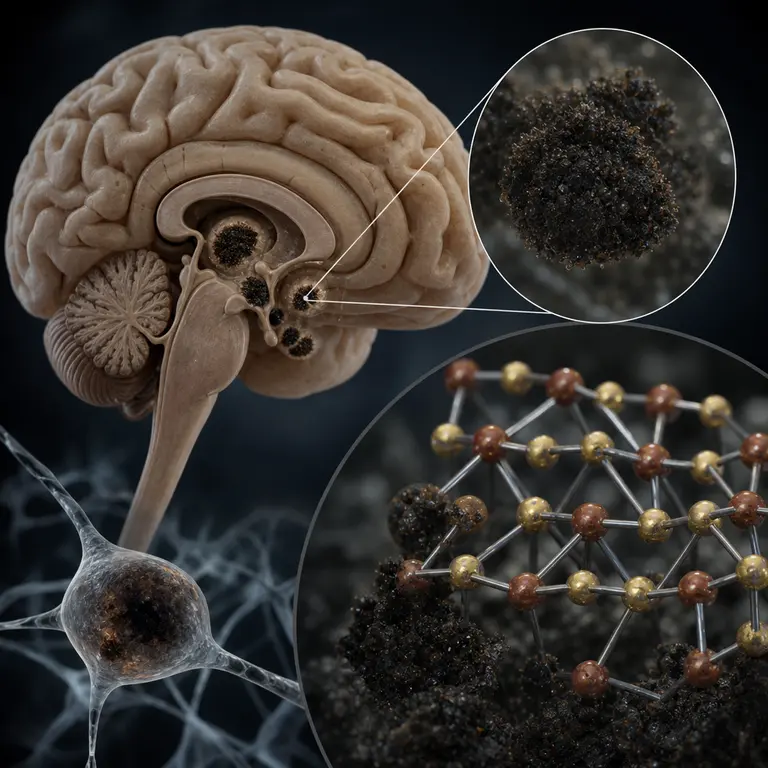
Deep within the human midbrain, in a crescent-shaped structure called the substantia nigra, lies a dark, granular pigment so unusual that it has no true equivalent in the mineral world outside the skull. Neuromelanin, the pigment responsible for the characteristic black coloration of this brain region, is not simply a biological dye. Over decades of a human lifetime, it accumulates iron and sulfur in a crystalline form that researchers have only recently begun to characterize with precision. A 2021 study published in JCI Insight, led by researchers at the University of Melbourne, used synchrotron X-ray fluorescence mapping to demonstrate that neuromelanin granules in aged human brains contain a specific iron-sulfur mineral phase resembling greigite — a magnetic iron sulfide more commonly associated with magnetotactic bacteria and ancient lake sediments — but structurally distinct enough to represent something genuinely novel in the biological world.
The substantia nigra, whose name is Latin for “black substance,” gets its color entirely from neuromelanin accumulation. In other primates, this region is far less darkly pigmented, and in most other mammals, it is essentially colorless. The degree of neuromelanin pigmentation in humans is uniquely pronounced, suggesting it is either a byproduct of our unusually high dopamine metabolism or serves some poorly understood neuroprotective function — or both. That a compound of this mineralogical strangeness sits quietly at the center of human neurochemistry, aging, and disease has only recently begun to receive the scientific attention it deserves.
How Neuromelanin Forms and Mineralizes Over Time
Neuromelanin is synthesized within dopaminergic and noradrenergic neurons via an oxidative pathway that involves excess cytosolic dopamine and norepinephrine. Unlike melanin in skin, which is produced by dedicated melanocyte cells and serves a clear photoprotective role, neuromelanin appears to form almost incidentally — as a kind of intracellular waste management system for reactive dopamine metabolites that would otherwise cause oxidative damage. The neurons of the substantia nigra produce dopamine in quantities that exceed what the cell can immediately package and deploy, and neuromelanin is, in part, what happens to the surplus.
What makes this pigment remarkable from a mineralogical standpoint is what happens as it ages. Neuromelanin granules act as chelating agents, binding iron from the cellular environment. In young brains, this iron is loosely associated with the organic polymer matrix of the pigment. But over decades, the iron undergoes a phase transformation. The 2021 Melbourne study, building on earlier synchrotron work by the same group, showed that in brains over 60 years old, the iron-sulfur core of neuromelanin granules transitions toward a more crystalline, magnetically ordered state. Iron loading in the substantia nigra of elderly humans can reach 200 to 400 micrograms per gram of wet tissue, among the highest levels found anywhere in the healthy brain.
This mineralization is not passive. The organic scaffold of neuromelanin appears to template the growth of these iron-sulfur clusters, in a manner analogous to how certain proteins in magnetotactic bacteria direct the formation of magnetite crystals. Magnetotactic bacteria are ancient microorganisms that navigate using Earth’s magnetic field by assembling chains of nanoscale magnetic minerals inside their cells, a process governed by highly specialized biomineralization proteins. The parallel with what neuromelanin does inside human neurons is striking, though the biological machinery behind this templating in the brain remains almost entirely uncharacterized. No dedicated protein complex for neuromelanin mineralization has yet been identified, which means the field does not yet know whether this process is controlled, incidental, or somewhere in between.
The granules themselves are not uniform structures. Electron microscopy has revealed that mature neuromelanin granules are composite bodies containing an organic melanin polymer core, lipid components, and the inorganic iron-sulfur mineral phase that accumulates with age. They are enclosed within lysosome-related organelles inside the neuron and appear to be largely stable over the lifetime of the cell. Because neurons in the substantia nigra are among the longest-lived cells in the human body — many surviving from birth to death without replacement — these granules have an unusually long time to accumulate material and undergo structural transformation. A neuromelanin granule in a 75-year-old brain has been mineralizing for seven decades.
The Parkinson’s Disease Connection
The reason this obscure neurochemistry matters urgently today is its intimate relationship with Parkinson’s disease, which affects more than 10 million people worldwide and whose incidence is rising faster than almost any other neurological condition. Parkinson’s disease is pathologically characterized by the selective death of dopaminergic neurons in the substantia nigra — precisely the cells that contain the most neuromelanin. As these cells die, the brain loses its capacity to produce dopamine in the quantities needed to coordinate smooth voluntary movement, leading to the tremor, rigidity, and bradykinesia that characterize the disease.
For decades, researchers debated whether neuromelanin was a victim or a contributor to this neuronal death. The emerging picture is complicated and in some ways troubling. When neurons die and rupture, they release neuromelanin granules into the extracellular space. These free granules, now loaded with their cargo of mineralized iron, trigger a powerful microglial inflammatory response. Microglia are the brain’s resident immune cells, and when they encounter extracellular neuromelanin, they mount an inflammatory reaction that can damage surrounding tissue. The iron released from degrading neuromelanin can also participate in Fenton chemistry, a well-characterized reaction in which ferrous iron reacts with hydrogen peroxide to generate hydroxyl radicals — among the most reactive and destructive oxidizing species in biology. In this way, neuromelanin may transform from a protective iron-sequestering system into a promoter of cell death as neurons containing it begin to fail. The very mechanism that kept neurons safe for decades becomes a liability at the moment of their death.
A 2023 paper in npj Parkinson’s Disease used neuromelanin-sensitive MRI — a relatively new imaging technique that exploits the paramagnetic properties of iron-laden neuromelanin — to show that substantia nigra neuromelanin signal loss precedes motor symptom onset by years in at-risk individuals. This finding is significant because by the time a Parkinson’s patient first notices a tremor, they have typically already lost more than half of their dopaminergic neurons in the substantia nigra. Any meaningful neuroprotective intervention would need to begin far earlier, which requires biomarkers capable of detecting the disease in its presymptomatic phase. Neuromelanin MRI is currently one of the most promising candidates for this role, with multicenter trials now underway in Europe, Japan, and North America. The fact that the imaging contrast depends on a mineralogical property of the pigment — its iron loading and magnetic character — rather than on any injected contrast agent makes it particularly attractive for longitudinal monitoring.
Evolutionary Implications and Open Questions
The evolutionary significance of such pronounced neuromelanin accumulation in humans remains genuinely puzzling. The substantia nigra of chimpanzees and gorillas, our closest relatives, shows only faint pigmentation compared to humans, despite their similarly dopamine-rich midbrain circuits. The divergence is not explained simply by brain size or overall dopamine production. Something about the human lineage specifically appears to have driven an unusual degree of neuromelanin accumulation, and the question of why has generated several competing hypotheses.
Some researchers have proposed that the intense neuromelanin pigmentation in humans is a metabolic consequence of the uniquely high prefrontal dopamine demands in humans. The cognitive load of language, abstract planning, long-horizon decision-making, and the management of complex social relationships may drive dopamine turnover rates in the human brain, thereby producing more neuromelanin as a byproduct. Under this view, the dark substantia nigra is essentially the metabolic exhaust of human cognition — an unavoidable consequence of the neurochemical infrastructure that makes complex thought possible.
Others have suggested a more provocative hypothesis: that neuromelanin’s iron-chelating capacity in humans evolved as a neuroprotective adaptation against dietary iron overload. The transition to meat-heavy diets in hominin evolution, which accelerated significantly with the controlled use of fire and the development of hunting technologies, would have substantially increased dietary heme iron intake. Free iron in the brain is neurotoxic, and a system capable of sequestering it within neurons rather than allowing it to circulate freely could have conferred a significant survival advantage. Under this view, the substantia nigra functions as a biological iron sink, protecting the rest of the brain from free iron toxicity — at the cost of making these neurons uniquely vulnerable to iron-mediated damage in old age, when the system eventually becomes overwhelmed.
These two hypotheses are not mutually exclusive, and the truth may involve elements of both. What they share is the suggestion that neuromelanin accumulation in humans represents not a flaw or an accident but an adaptation that carried real benefits during the reproductive years of our evolutionary history, while imposing costs that manifest primarily in post-reproductive aging. This is a pattern familiar from evolutionary medicine more broadly — the concept of antagonistic pleiotropy, in which traits selected for early-life benefit become liabilities in old age.
Conclusion
Neuromelanin sits at an extraordinary intersection of mineralogy, evolutionary biology, and clinical neuroscience. It forms a compound inside human neurons that geologists have no established name for, accumulates over a lifetime in a process that mirrors biomineralization in ancient microorganisms, and when it fails, contributes to one of the most debilitating diseases of modern aging. It is a structure that has existed in every human brain since before our species had a name for the color black, yet its precise chemistry was not characterized until powerful synchrotron light sources capable of resolving nanoscale mineral phases in biological tissue became available in the twenty-first century.
The story of neuromelanin is also a reminder of how much remains unknown about the basic material composition of the human brain. Decades of neuroscience focused on electrical signaling, neurotransmitter dynamics, and synaptic plasticity have produced extraordinary knowledge, but they have left relatively unexplored the question of what the brain is made of at the mineralogical level. The answer, at least in the substantia nigra, turns out to be something that does not fit neatly into any existing category — not quite geology, not quite biochemistry, but a slow crystallization of human experience into matter. The mineral inside your brain may, in a very literal sense, be unique to the human condition.
Sources & Further Reading
- Zucca FA, Segura-Aguilar J, Ferrari E, et al. Interactions of iron, dopamine and neuromelanin pathways in brain aging and Parkinson's disease. Progress in Neurobiology, 2017. https://doi.org/10.1016/j.pneurobio.2015.09.012
- Kostka JK, Tebo BM, et al. (Melbourne group, Hare D). Synchrotron X-ray fluorescence mapping of neuromelanin iron mineralogy in the human substantia nigra. JCI Insight, 2021. https://insight.jci.org
- Fujita K, Sato S, Watanabe M, et al. Neuromelanin-sensitive MRI as a biomarker for Parkinson's disease progression. npj Parkinson's Disease, 2023. https://doi.org/10.1038/s41531-023-00484-4
- Double KL. Neuromelanin in human substantia nigra: an overview of its neurochemical characteristics. Neurotoxicity Research, 2006.