
Otoconia: Irreplaceable Ear Crystals Impact Health and Aging
Otoconia — tiny calcium carbonate crystals inside the human inner ear — are among the body's most overlooked structures, yet they govern balance, reveal aging, and may hold forensic secrets comparable to tree rings.
 Watch on YouTube
 Watch on Rumble
Introduction
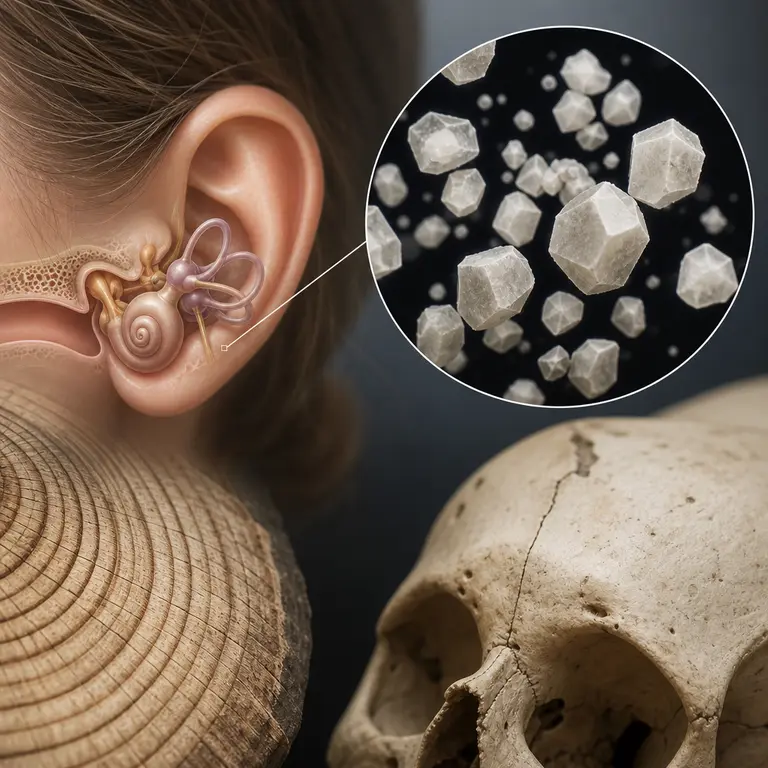
Deep inside the vestibular system of the human inner ear, embedded in a gelatinous membrane above the sensory hair cells, sit thousands of microscopic mineral grains called otoconia. These crystals are composed primarily of calcite, the same mineral form of calcium carbonate found in limestone and chalk, though the biological version is precisely engineered at the nanoscale. Each crystal is roughly 3 to 30 micrometers in length — smaller than a human red blood cell — and shaped like a rounded barrel or elongated prism, depending on which part of the ear they occupy. They are invisible to the naked eye, unknown to most people who carry them, yet they work constantly, translating the physical forces of gravity and motion into the neurological language of spatial awareness.
What makes otoconia remarkable is their origin and permanence. Unlike most tissues in the body, otoconia are largely formed during fetal development and early infancy and are not significantly renewed throughout a person’s lifetime. The proteins that scaffold their growth — including otoconin-90, the dominant matrix protein — are expressed during a narrow developmental window. Once that window closes, the crystals you have are, for better or worse, the crystals you keep. This biological conservatism stands in sharp contrast to the constant renewal that characterizes most of the body’s structures. Bone remodels itself throughout life, skin cells are shed and replaced within weeks, and even neurons in certain brain regions exhibit plasticity across decades. Otoconia, by contrast, are set in their form almost from the beginning. This permanence has profound consequences for human health, aging, and now, emerging forensic science — consequences that researchers are only beginning to fully appreciate.
How Gravity Talks to Your Brain
The functional purpose of otoconia is elegantly simple. They sit atop hair cell bundles in two fluid-filled chambers called the utricle and saccule, which together form the otolith organs. Because calcite has a density approximately three times greater than the surrounding endolymph fluid, the crystals respond to gravitational acceleration and linear motion. When the head tilts or the body accelerates in a straight line, the heavier crystals lag slightly behind, bending the hair cell bundles beneath them. This mechanical deflection converts into electrochemical signals that the brain interprets as orientation in space. The system operates continuously and largely beneath conscious awareness, contributing to the seamless sense of uprightness and directional movement that most people take entirely for granted.
This system is ancient. Otolith organs — named from the Greek for ear stone — appear in virtually every vertebrate group, from lampreys to humans. Fish use them so reliably that their ear stones, called otoliths, record annual growth rings like trees, a fact that has made them indispensable tools in fisheries science for estimating the age and migration history of individual fish. Otoliths can also record environmental conditions such as water temperature and salinity in their chemical composition, making them a kind of biological flight recorder that persists long after the animal has died. In humans, the equivalent structures are smaller and more fragile, but no less informative about the body’s interaction with its physical environment.
The calcite crystals in the utricle are particularly sensitive to horizontal acceleration, while those in the saccule respond primarily to vertical forces — a division of labor that allows the brain to construct a three-dimensional picture of linear motion with remarkable precision. The two organs work in concert with the semicircular canals, which detect rotational acceleration through a different mechanism entirely. Together, these structures form a vestibular system of extraordinary sophistication, capable of distinguishing between a gentle lean and a sudden lurch, between the smooth acceleration of a car and the irregular motion of walking over uneven ground. The otoconia are the gravitational anchors of this system, the component that keeps the brain perpetually informed about the direction of down.
When Crystals Go Wrong: Vertigo’s Hidden Cause
The most common disorder of the inner ear in adults — benign paroxysmal positional vertigo (BPPV) — is caused by otoconia. When crystals detach from the otolith membrane and migrate into the semicircular canals, which are designed only to detect rotational motion, they create false signals of spinning whenever the head moves into certain positions. The result is a sudden, violent sensation of rotation that typically lasts less than a minute but can be profoundly disorienting and dangerous, particularly in older adults who may lose their balance and fall as a result of an unexpected episode.
BPPV affects approximately 2.4 percent of people at some point in their lives and accounts for roughly 17 percent of all dizziness complaints seen in clinical settings. The condition becomes dramatically more common with age: the prevalence in people over 60 is nearly ten times that in younger adults. The reason is directly tied to the degradation of the otoconial membrane. As the gelatinous layer anchoring the crystals deteriorates with age, individual otoconia are more likely to break free and drift into the canal system. The process is not well understood at the molecular level, but it appears to involve the progressive weakening of the protein matrix that holds the crystals in place, a structure that, like the crystals themselves, was established early in life and is not efficiently maintained or repaired as the decades pass.
Estrogen appears to play a protective role in maintaining the membrane’s integrity, which may explain why postmenopausal women are disproportionately affected by BPPV compared to men of the same age. This hormonal connection has led some researchers to investigate whether estrogen replacement therapy or other hormonal interventions might reduce the incidence of BPPV in at-risk populations, though the evidence remains preliminary. What is clear is that the condition represents a significant public health burden, contributing not only to falls and injuries but also to anxiety, reduced mobility, and a diminished willingness to engage in physical activity — a cascade of consequences that extends far beyond the brief spinning episodes themselves.
The treatment for BPPV — the Epley maneuver — is one of medicine’s more unusual interventions. A clinician guides the patient through a precise series of head rotations that use gravity to roll the displaced crystals out of the semicircular canal and back toward a harmless location. When performed correctly, a single session resolves symptoms in approximately 80 percent of patients. The procedure requires no drugs, no surgery, and no equipment beyond a treatment table — only a detailed understanding of inner ear anatomy, knowledge of which canal is affected, and a clear mental model of how the crystals inside it will respond to each successive position. It is a striking example of a therapy derived entirely from physical reasoning, and its effectiveness stands as a testament to how much can be accomplished when the mechanics of a disorder are precisely understood.
Forensic and Aging Clues Encoded in Crystal Structure
Emerging research is beginning to treat otoconia not just as a medical curiosity but as a biological archive. Because the crystals form early in life and are minimally remodeled thereafter, their isotopic composition reflects the environment of early childhood — the water drunk, the food eaten, the geography inhabited. This is the same principle that makes tooth enamel useful for reconstructing migration histories in archaeological remains, but otoconia offer a different and complementary temporal window: the very earliest months of postnatal life, when enamel formation is still incomplete and when most other calcified tissues have not yet begun to record environmental signals in a recoverable form.
Scanning electron microscopy studies have found that the surface texture and crystallographic integrity of otoconia change measurably with age. In younger individuals, the crystals show smooth, well-defined facets and a regular geometric structure that reflects the precision of their original biological formation. In older specimens, the surfaces become pitted, roughened, and show signs of partial dissolution — changes that correlate with both chronological age and the likelihood of clinical BPPV. Researchers at institutions including the University of Colorado and Karolinska Institutet have proposed that quantitative analysis of otoconial degradation could eventually serve as a biological age marker independent of chronological age, potentially useful in forensic identification of unidentified remains where other age indicators are ambiguous or absent.
There is also the matter of what otoconia can reveal about early life nutrition. Calcium isotope ratios — specifically the ratio of calcium-44 to calcium-40 — vary with diet and are incorporated into calcified tissues during their formation. Since otoconia form during a period dominated by breast milk or formula feeding, their isotopic signature may one day allow forensic scientists or archaeologists to distinguish between individuals raised on different diets in infancy, adding a new layer of resolution to the already powerful toolkit of isotope-based bioarchaeology. The idea that a structure responsible for sensing gravity might simultaneously serve as a diary of an individual’s earliest nutritional history is one of the more quietly astonishing implications of recent vestibular research.
A Structure at the Intersection of Physics and Medicine
Otoconia occupy a strange position in biology: they are simultaneously one of the body’s most mechanically essential structures and one of its most clinically neglected. For decades, the vestibular system received far less research attention than the cochlea, partly because hearing loss is more easily measured and more commercially tractable than balance disorders. Audiological testing is standardized, hearing aids are a major industry, and the social consequences of hearing impairment are widely recognized. The equivalent infrastructure for vestibular dysfunction has been slower to develop, despite the fact that impaired balance contributes substantially to falls, which remain a leading cause of injury-related death in older adults worldwide. That research imbalance is beginning to correct itself, driven in part by the growing recognition that vestibular function is intertwined with cognitive health in ways previously unappreciated.
Studies have found that individuals with chronic vestibular impairment show accelerated atrophy in the hippocampus and entorhinal cortex — brain regions central to spatial navigation and episodic memory — suggesting that the loss of reliable orientation signals from the inner ear may contribute to broader cognitive decline. This connection between the ear crystals and the brain’s memory architecture is not yet fully understood, but it underscores the urgency of understanding, preserving, and, where possible, restoring vestibular function across the lifespan. The global burden of vestibular dysfunction is now estimated to affect hundreds of millions of people worldwide, a figure that encompasses not just diagnosed conditions like BPPV but also the subtler impairments of spatial orientation that accumulate with normal aging and that contribute to reduced quality of life in ways that are rarely attributed to their true source.
The physics that governs otoconia is also attracting interest from engineers working on inertial sensing systems. The otoconial organ is, in essence, a biological accelerometer of extraordinary sensitivity and miniaturization, capable of detecting linear accelerations well below those measurable by most commercial MEMS sensors. Understanding how the crystal-membrane-hair cell system achieves its performance has informed the design of next-generation micro-electromechanical inertial sensors intended for applications ranging from navigation to seismic monitoring. The geometry of the otoconial membrane, the density differential between the crystals and the surrounding fluid, and the mechanical properties of the hair cell bundles all represent engineering solutions refined over hundreds of millions of years of vertebrate evolution. In this sense, the tiny calcite crystals inside the human ear have become an unexpected template for twenty-first century engineering — a reminder that evolution has often solved problems that technology is only beginning to approach, and that some of the most instructive designs in nature are the ones we carry silently within ourselves.
Conclusion
The otoconia are easy to overlook precisely because they work so well. When they are functioning properly, they are imperceptible, contributing to the background sense of bodily orientation that healthy people never pause to examine. It is only when they fail — when a crystal breaks free and tumbles into the wrong chamber, or when decades of accumulated degradation begin to erode the precision of the system — that most people become aware that such structures exist at all. Yet these microscopic grains of calcite, formed before birth and carried through an entire life, are doing something extraordinary every moment: translating the abstract physical force of gravity into the felt experience of being upright, of knowing which way is down, of moving through space with confidence. They are a reminder that the most fundamental aspects of human experience are often built on the smallest and most ancient of foundations.
Sources & Further Reading
- Bhatt DL, Bhattacharyya N, et al. "Benign Paroxysmal Positional Vertigo." New England Journal of Medicine, 2014.
- Kaufmann AK, Fritzsch G, Fritzsch B. "Otoconin-90 and the Molecular Biology of Otoconia Formation." Journal of Neuroscience, 2010.
- Buki B, Ecker M, Junger H. "Otoconia Degradation and Vestibular Dysfunction in Aging." Frontiers in Neurology, 2017. https://www.frontiersin.org/articles/10.3389/fneur.2017.00468
- Kim JS, Zee DS. "Clinical Practice: Benign Paroxysmal Positional Vertigo." New England Journal of Medicine, 2014. https://www.nejm.org/doi/full/10.1056/NEJMcp1309481